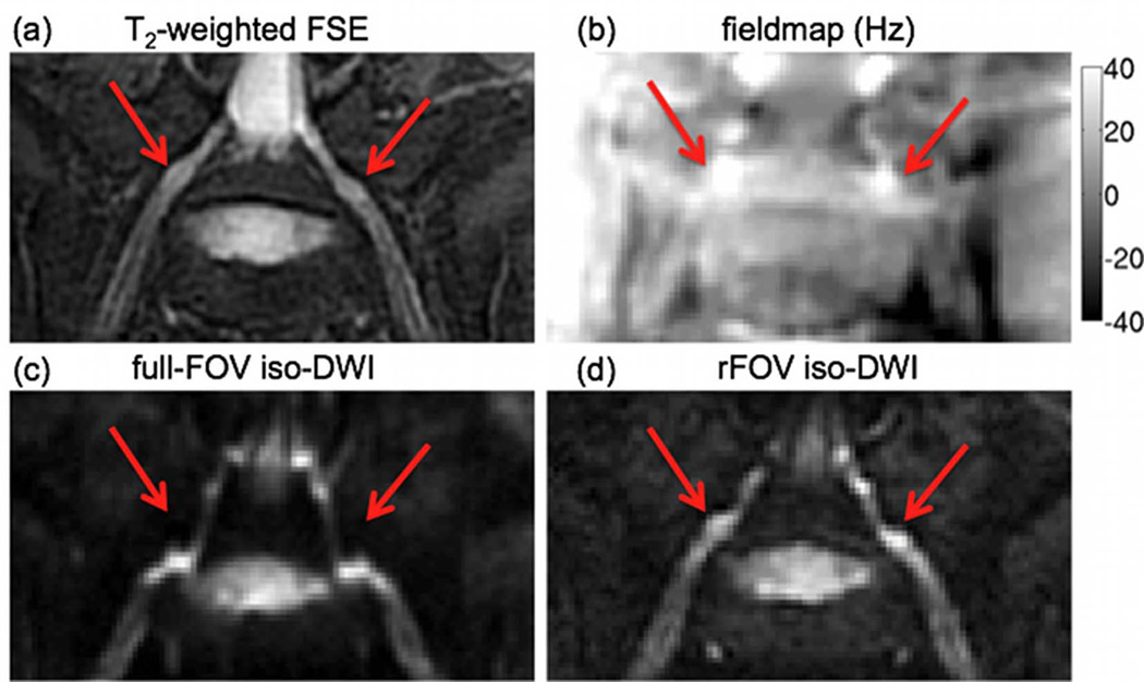
Figure 3.
Effect of geometric distortions: (a) oblique coronal T2-weighted FSE, (b) fieldmap (grayscale map values in Hz) and oblique coronal iso-diffusion weighted images using (c) full-FOV EPI and (d) reduced-FOV EPI. The red arrow points to the intervertebral foraminal region showing strong fieldmap variations and significant geometric distortions in the full-FOV EPI acquisition along the PE direction (S/I direction).

