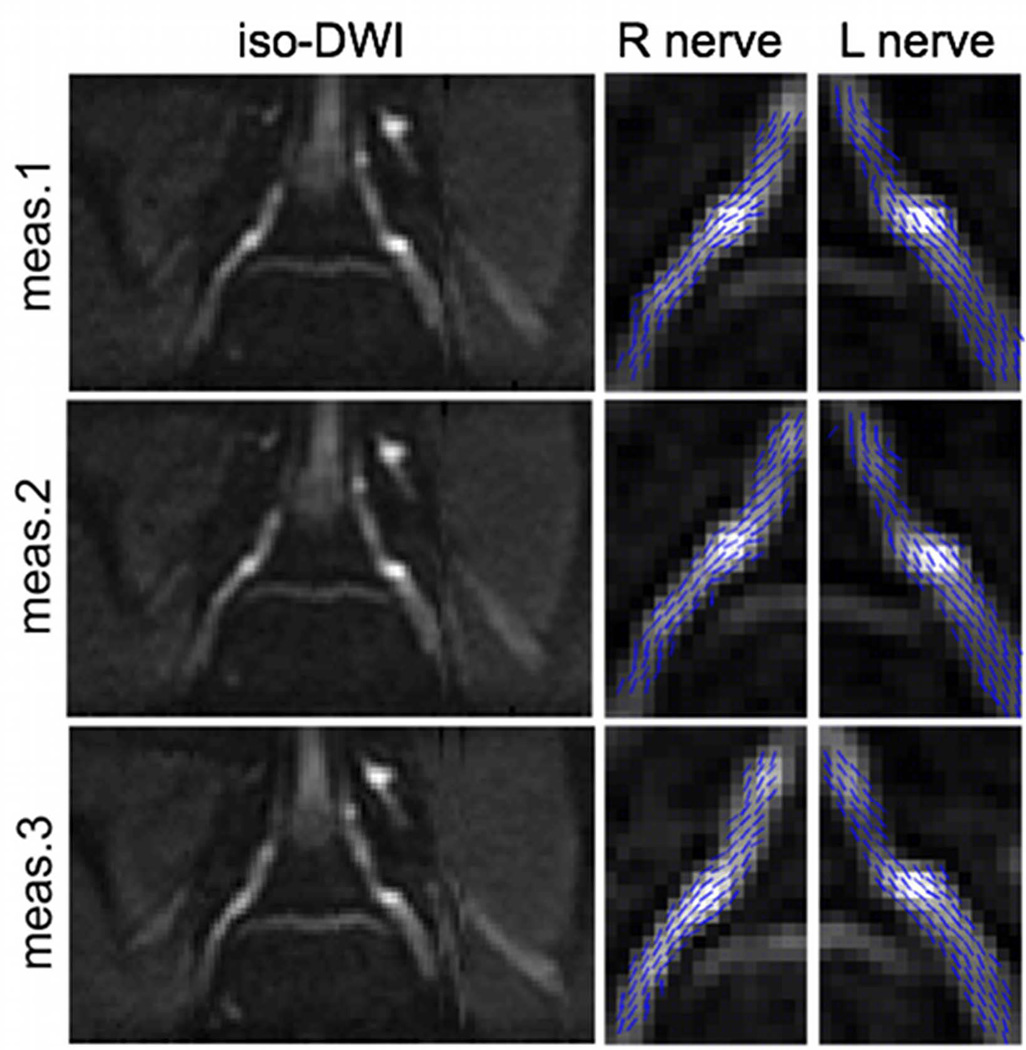
Figure 5.
Repeatability of L5 nerve root DTI of the same subject at three different exams. First column shows the bilateral iso-diffusion weighted images. Second column shows the right nerve primary diffusion tensor eigenvector projections on the slice plane superimposed on iso-diffusion weighted images. Third column shows the left nerve primary diffusion tensor eigenvector projections on the slice plane superimposed on iso-diffusion weighted images.

