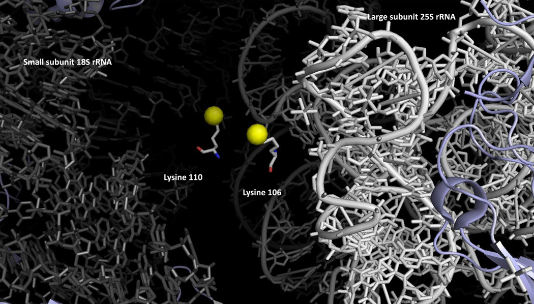
Figure 3.
Intrasubunit localization of the methylated lysine residues of Rpl23ab in yeast cytoplasmic ribosomes. Dimethyllysine residues 105 and 109 are shown with the epsilon amino group as a yellow sphere. These residues are positioned at the interface of the small and large ribosomal subunits; 18S rRNA is shown in gray on the left and 25S rRNA in white on the right. The illustration was made using PyMOL from the PDB structures 3U5F, 3U5G, 3U5H, and 3U5I [45].

