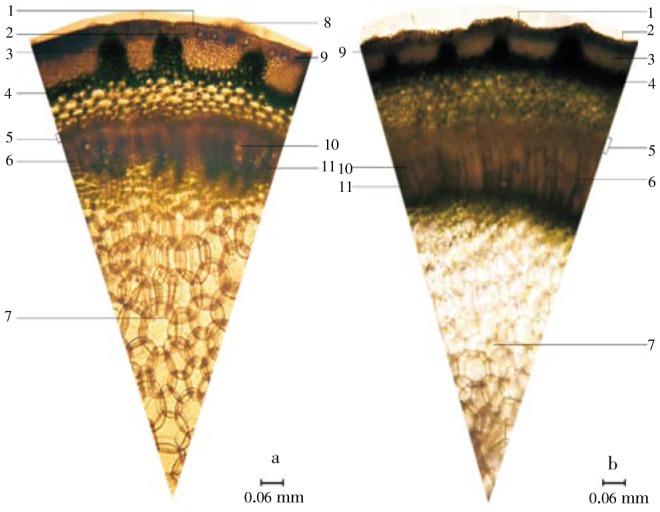
Figure 3. Stem cross section of C. nutans and C. siamensis.

a: C. nutans; b: C. siamensis; 1: epidermis; 2: lithocyst; 3: group of collenchyma; 4: parenchyma of cortex; 5: phloem tissues; 6: parenchyma ray; 7: ground tissue; 8: multicellular trichome; 9: cortical parenchyma; 10: xylem vessel; 11: xylem fiber.
