Fig. 1.

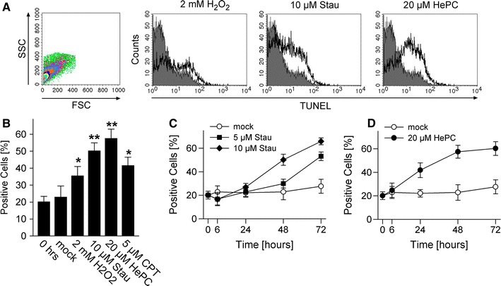
Pro-apoptotic stimuli trigger DNA fragmentation in T. gondii. Parasites were treated with hydrogen peroxide (H2O2), staurosporine (Stau), miltefosine (HePC) or camptothecin (CPT) as indicated, or were mock-treated. At different time points after treatment, DNA strand breaks were visualized by in situ TUNEL labelling and parasites were analysed by flow cytometry. Freshly isolated parasites were processed in parallel (0 h). a Parasites were selected according to FSC/SSC parameters and were analysed for terminal deoxynucleotidyl transferase-mediated dUTP nick end labelling (TUNEL). The graphs show representative histograms from parasites which have been treated with the indicated stimuli for 48 h (open histograms), or those which have been freshly isolated (filled histograms). b Mean percentages ± SEM of TUNEL-positive T. gondii as determined by flow cytometry; results are from 5 to 6 independent experiments (CPT: n = 2). Significant differences between treated T. gondii and untreated controls are indicated by one (p < 0.05) or two asterisks (p < 0.01). c, d Kinetics of DNA fragmentation in T. gondii following treatment with staurosporine or miltefosine or following mock treatment. Data represents mean ± SEM from at least three independent experiments
