Fig. 5.

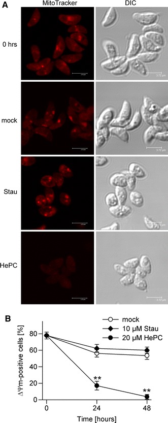
Loss of mitochondrial membrane potential (ΔΨ m) during pro-apoptotic treatment of T. gondii with miltefosine. Parasites were treated with 10 μM staurosprine (Stau) or 20 μM miltefosine (HePC), or were mock-treated for 24 and 48 h. Freshly isolated parasites and treated parasites were incubated with MitoTracker probe Orange CM-H2TMRos. After fixation, cells were analysed by confocal laser scanning microscopy. a Representative images from three independent experiments are depicted. b The percentages of cells with an intact ΔΨ m were determined by counting at least 500 parasites per sample; data represents mean ± SEM from three independent experiments. Significant differences are indicated (**p < 0.01)
