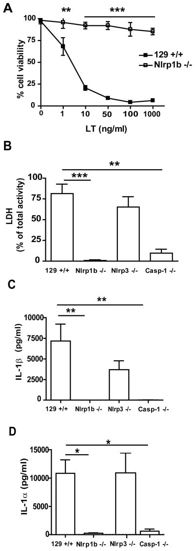
Figure 2. Lethal toxin mediated cell death and IL-1β and IL-1α cytokine release from macrophages.
(A) BMDMs from 129+/+ and Nlrp1b −/− mice were incubated with LT for 4 hours and cell viability was determined with the WST-1 reagent. (B–D) Thioglycolate-elicited peritoneal macrophages were primed with 100 ng/ml ultra-pure LPS overnight and subsequently incubated for 6 hours in presence of 1 μg/ml LT. Cell death was determined by LDH activity in supernatant and expressed as the percentage of total cellular LDH activity (B). IL-1β (C) and IL-1α (D) release to supernatant was detected by ELISA. n=4 for 129+/+, Nlrp1b −/−, Nlrp3 −/−; n=3 for Casp-1 −/−. * p<0.05, ** p<0.01, *** p<0.001

