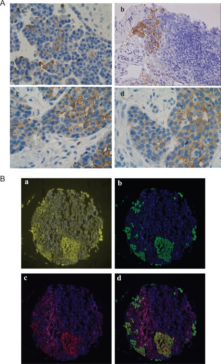
Figure 1.
Expression of CK-10 and CD44 in ovarian cancer. A, Representative immunostaining for CK19 in ovarian cancer samples. B, Representative examples for CD44 and CD44/CK19 staining patterns in Yale ovarian cancer 203 TMA. 4′,6-Diamidino-2-phenylindole ([DAPI] nucleus) is blue, pan-cytokeratin is yellow, CK19 is green, and CD44 is red. (a) Pan-cytokeratin and DAPI staining. (b) CD44 and DAPI staining. (c) CK19 and DAPI staining. (d) CD44, CK19, and DAPI staining merged.

