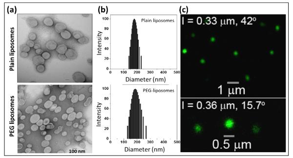
Figure 5.
Physicochemical characterization of liposomes: (a) Transmission electron micrographs, (b) Dynamic light scattering for particle size distribution, and (c) One-photon Laser Scanning microscopic images of HDAS-PEG liposomes. The sample was stained with Alexa Fluor® 488 conjugated donkey anti-goat IgG antibody. The emission was collected at 500-700 nm. The wavelength for the excitation was 495 nm.

