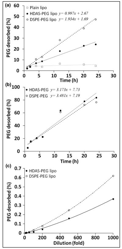
Figure 6.
(a) HDAS-PEG and DSPE-PEG desorption (with respect to time) after its insertion into the preformed liposomes. A 7 KDa MWCO dialysis cassette was used to separate desorbed PEG-lipids (MW ~2,500) from the PEGylated liposomes. No PEG was detected in the dialysates from plain liposomes (open squares). (b) The micellar solutions of HDAS-PEG and DSPE-PEG (1 mM) served as positive controls in the assay. A linear release of the majority of PEG-lipids confirms the ability of dialysis

