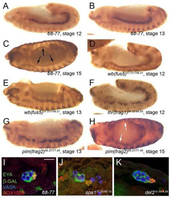
Figure 3. Visceral mesoderm and msSGP phenotypes observed in gonad mutants.
(A-H) Visceral mesoderm phenotypes. Embryos immunolabeled for the visceral mesoderm marker FASIII (anti-FASIII), stages and genotypes indicated. (A-C) In wild type the visceral mesoderm is a smooth, coherent tissue at stages 12 (A) and 13 (B), and has surrounded the gut by stage 15 (black arrows, C). (D, E) The visceral mesoderm in wb(fus5) mutants is fused into a coherent tissue at both stage 12 (D) and 13 (E), but is thinner than normal, and jagged in appearance. (F-H) The visceral mesoderm in thr(frag1) (F) and pim(frag2) (G, H) mutants does not completely fuse, leaving breaks in the tissue. At younger stages (F, G), some visceral mesoderm clusters appear to be contacting each other but have not integrated together. (H) The visceral mesoderm is able to surround the gut by stage 15 in pim(frag2) mutants, but several thin spots or even holes are seen in the tissue (white arrows). (I-K) Examples of msSGP phenotypes. Embryos immunolabeled for SGPs (68-77 enhancer trap, anti-β-Galactosidase, and anti-EYA, both green), msSGPs (anti-SOX100B, red and anti-EYA, green – appear yellow) and GCs (anti-VASA, blue). Stage 15 embryos, all male. Bar = 20μm. (I) Wild type embryo. msSGPs have joined and integrated into the gonad. (J) A gonad whose msSGPs failed to join (spa1). (K) A gonad with msSGPs that have joined but are loosely gathered (del2).

