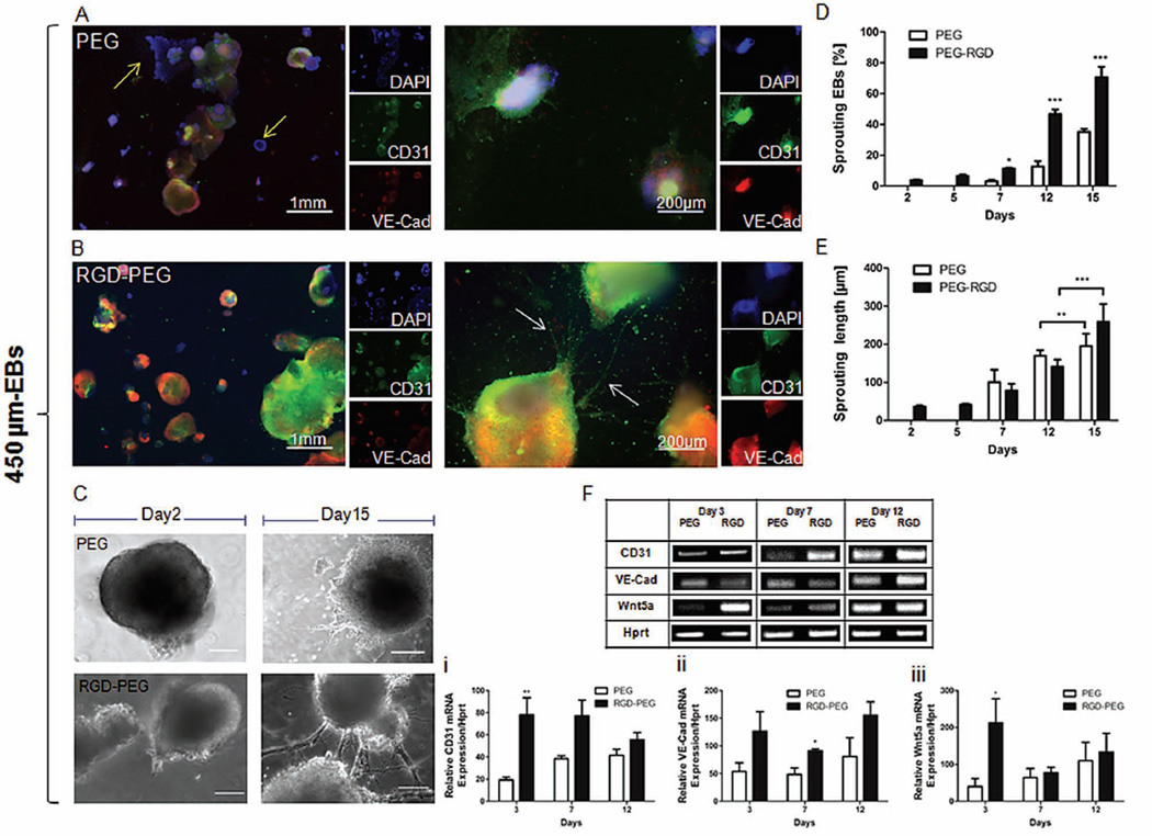
Figure 4.
Enhanced endothelialization in 450 µm-EBs with RGD-conjugation. (A) Immunocytochemical characterization of vasculogenic markers (CD31 or VE-Cadherin) in unmodified PEG polymer (Arrow indicates CD31- and VE-Cadherin- unstained areas) and (B) in RGD-PEG (Arrows indicate sprouting areas). (C) Phase contrast microscopic images of encapsulated EBs in PEG at day 2 (no sprouting visualized) and day 15. Sprouting in RGD-PEG started at day 2 of encapsulation (5 days earlier than in unmodified PEG). (Scale bars for day 2: 100 µm, day 15: 200 µm). (D)% of sprouting EBs with initial diameter size of 450 µm. The number of sprouting EBs was counted in time course and normalized to the number of encapsulated EBs. (E) Length measurements of sprouts were performed with imageJ. Values refer to the development of the length per one sprout and EB. Starting by the following time point, sprouts with a length less than 100 µm were taken out of consideration. Sprouting was visualized with phase contrast microscopy. (F) Gene expression analysis on 450 µm-EBs in PEG vs. RGD-PEG; Housekeeping gene (Hprt) and endothelial cell markers (CD31, Wnt5a, and VE-Cadherin) were screened on 450 µm-EBs at day 3, day 7, and day 12 of encapsulation (n = 3, * indicates P ≤ 0.05 compared to RGD-PEG). Error bars without * do not represent statistical significance.

