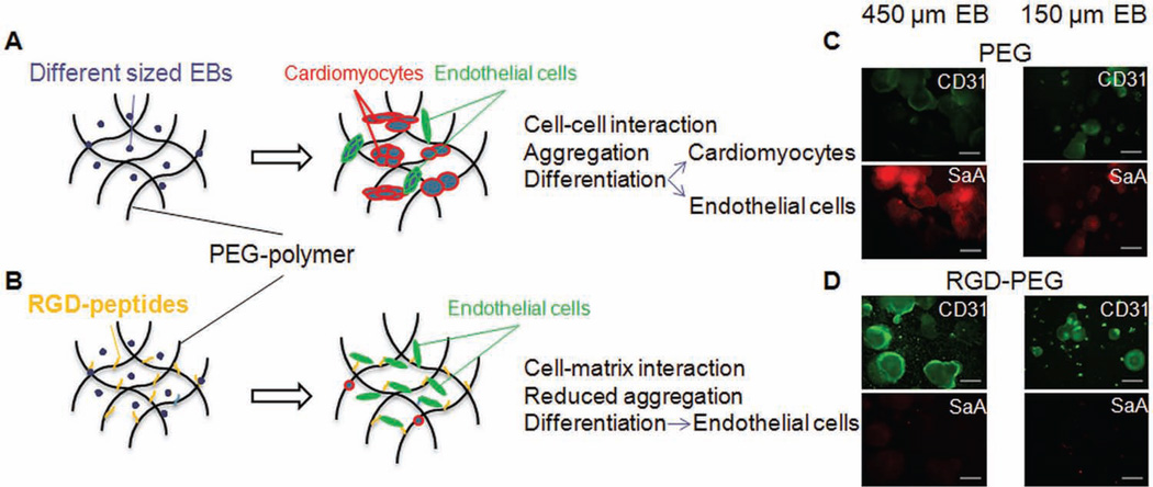
Figure 6.
(A) Schematic structure of PEG polymer containing EBs of different sizes. After culturing samples in PEG polymer, aggregation was visualized in both EB-sizes. Immunocytochemical-, gene expression-, and quantification data showed that EBs in PEG can differentiate towards cardiomyocytes and endothelial cells. (B) RGD-peptides were conjugated in PEG polymer. EBs in RGD-PEG showed lower aggregation, by increased differentiation towards endothelial cells and decreased differentiation towards cardiomyocytes. (C) Immunocytochemical staining of EBs (450 µm left and 150 µm right) encapsulated in PEG polymer. EBs expressed cardiogenic marker Sacromeric alpha actinin and endothelial marker CD31. (D) Different sized EBs embedded in RGD-PEG showed higher expression of CD31 and a significant decrease of Sacromeric alpha actinin expression. Scale bars (C, D): 500 µm.

