Fig. 6.
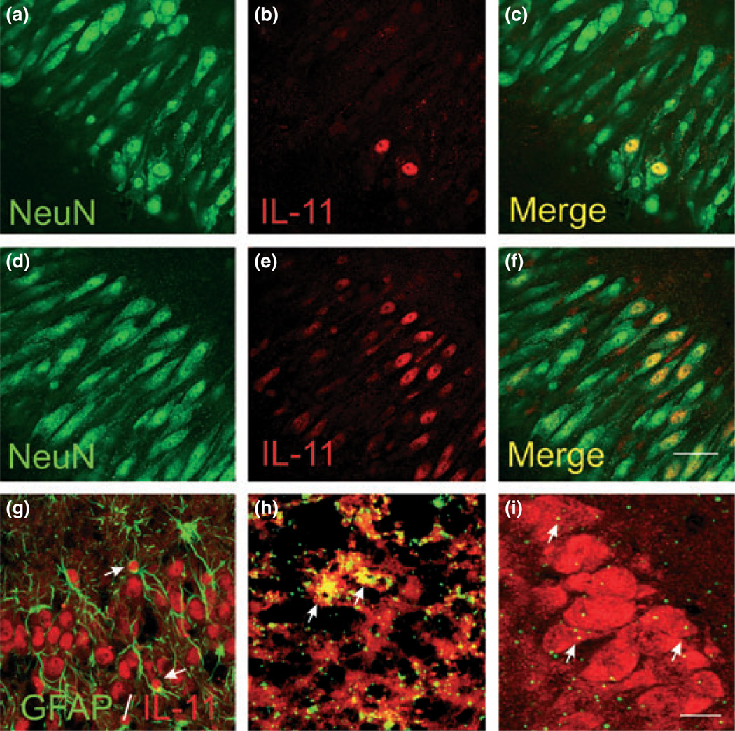
Cellular localization of cold pre-conditioning TNF-α and IL-11 changes. (a–c) Control hippocampal slice culture immunostaining with neuronal marker NeuN (a) followed by IL-11 immunostaining (b) showed that the predominant cell type positive for IL-11 immunoreactivity were pyramidal neurons (c). (d–f) Exposure to TNF-α (100 ng/ mL) for 24 h dramatically increased neuron-specific IL-11 immunostaining. Here, we show exemplary confocal photomicrographs (n = 5 per group). Scale bar, 50 µm. (g) Image shows double-labeling with glial fibrillary acidic protein (GFAP) and IL-11 immunostaining at the pyramidal cell layer from an exemplary hippocampal slice culture 24 h after cold pre-conditioning (30°C for 90 min). We observed IL-11-immunopositive astrocytes (arrows) in only a minority of photomicrographs (n = 5). (h) Image shows that mRNA for TNF-α (green) 3 h after cold pre-conditioning (90 min at 30°C) localized to microglia, immunostained with CD11b (red). The resultant exemplary (from n = 5) image shows green spots overlying red cells or the optical conversion of these spots to yellow when both in situ and immunostaining probes are blended at the same plane of focus (arrows). (i) Twelve hours after analogous cold pre-conditioning (n = 5), mRNA for IL-11 (green) localized mainly to pyramidal neurons immunostained with NeuN (red). Arrows point to exemplary in-focus in situ and immunostaining probe markings. Although sparse, these probe markings for IL-11 mRNA concentrate to the pyramidal neuron layer. (g–i) Scale bar, 25 µm (g and h) and 50 µm (i).

