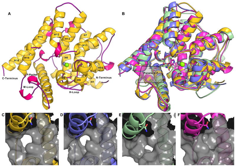
Figure 2.
(A) An overview of the X-ray crystal structure of TbrPDEB1, helices are colored yellow and labeled where visible, 310-helices are shown in fuchsia, and loops are shown in purple. The metal ions magnesium (lime) and zinc (gray) are shown. (B) A superposition of crystal structures of TbrPDEB1 (yellow), LmjPDEB1-IBMX (2R8Q in blue)12, TcrPDEC-WYQ16 (3V94 in green)4, and a crystal structure of hPDE4B-piclamilast (1XM4 in magenta).17 Close-ups of the region between Q874 on H15 and the M-loop are shown in: (C) TbrPDEB1; (D) LmjPDEB1; (E) TcrPDEC; and (F) hPDE4B.

