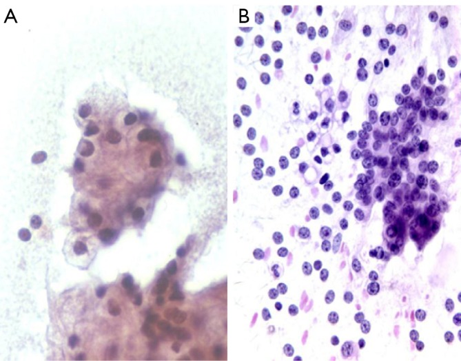
Figure 10.

A. acinar cell carcinoma with solid overlapping nested cluster of large cells with granular cytoplasm and round nuclei (Pap stain, 400×); B. acinar cell carcinoma with numerous stripped nuclei in the background (DQ stain, 200×)

A. acinar cell carcinoma with solid overlapping nested cluster of large cells with granular cytoplasm and round nuclei (Pap stain, 400×); B. acinar cell carcinoma with numerous stripped nuclei in the background (DQ stain, 200×)