Figure 13.

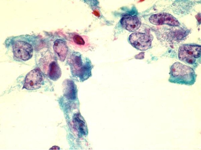
Adenosquamous carcinoma, showing occasional squamoid tumor cells with orangeophilic dense cytoplasm with distinct cell borders as well as glandular tumor cells with hypochromatic nuclei and prominent nucleoli (Pap stain, 400×)

Adenosquamous carcinoma, showing occasional squamoid tumor cells with orangeophilic dense cytoplasm with distinct cell borders as well as glandular tumor cells with hypochromatic nuclei and prominent nucleoli (Pap stain, 400×)