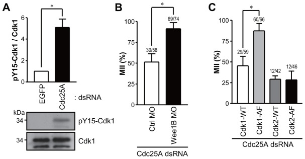
Fig. 3.

Cdk1 phosphorylation in Cdc25A knockdown oocytes. (A) Immunoblot analysis of phospho-tyrosine Cdk1 in Cdc25A knockdown oocytes. MII oocytes injected with EGFP or Cdc25A dsRNA were incubated for 24 hours and subjected to SDS-PAGE followed by immunoblotting for Cdk1 and Cdk1 phospho-tyrosine 15 (pY15-Cdk1). The pY15-Cdk1/Cdk1 ratio is reported together with an immunoblot representative of three experiments performed. (B) In vitro matured MII oocytes injected with control (Ctrl) or Wee1B MO were injected with Cdc25A dsRNA and incubated for 24 hours. (C) Cdc25A dsRNA was co-injected into MII oocytes with mRNAs encoding Cdk1-WT, Cdk1-AF, or Cdk2-WT and Cdk2-AF. The percentage of treated oocytes in MII was determined for B and C. Data are the means ± s.e.m. of at least three independent experiments. Numbers above the bars indicate the number of MII oocytes/total oocyte number. *P<0.05.
