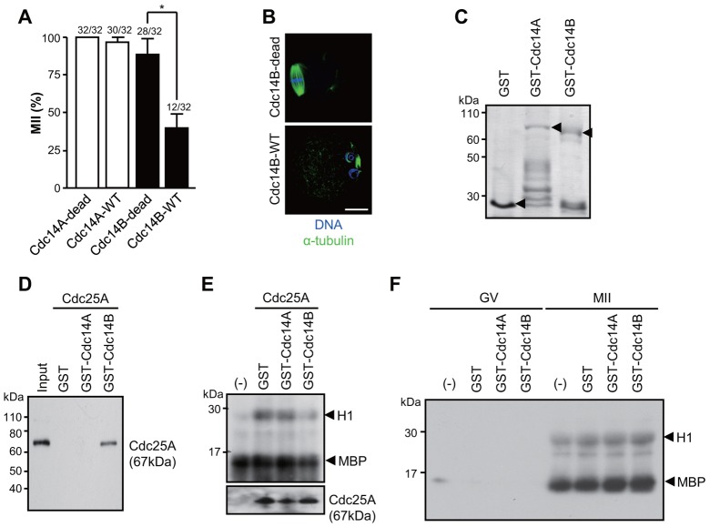
Fig. 4.
Cdc14 effect on MII arrest and interaction with Cdc25A. (A) MII oocytes were injected with mRNAs coding for Cdc14A or B, and the percentage of treated oocytes in MII was scored after 24 hours. Numbers above the bars indicate the number of oocytes in the MII stage as well as total number of oocytes used. *P<0.005. (B) Representative images of Cdc14B-overexpressing oocytes. The spindle and DNA were stained with anti-α-tubulin and DAPI, respectively. Scale bar: 20 µm. (C) GST fusion proteins used in the pull-down assay (arrowhead) were separated by SDS-PAGE and stained with Coomassie Brilliant Blue. (D) GST fusion proteins were immunoprecipitated from the cell lysates overexpressing Cdc25A. Proteins recovered in the immunoprecipitation pellet were immunoblotted for Cdc25A. (E) Immunoprecipitated Cdc25As were pre-incubated with GST fusion proteins and H1/MBP double kinase assay was performed with GV oocyte lysates. (F) GST fusion proteins were subjected to H1/MBP double kinase assay with GV or MII oocytes lysates. Water was loaded as a negative control (−).

