Fig. 5.
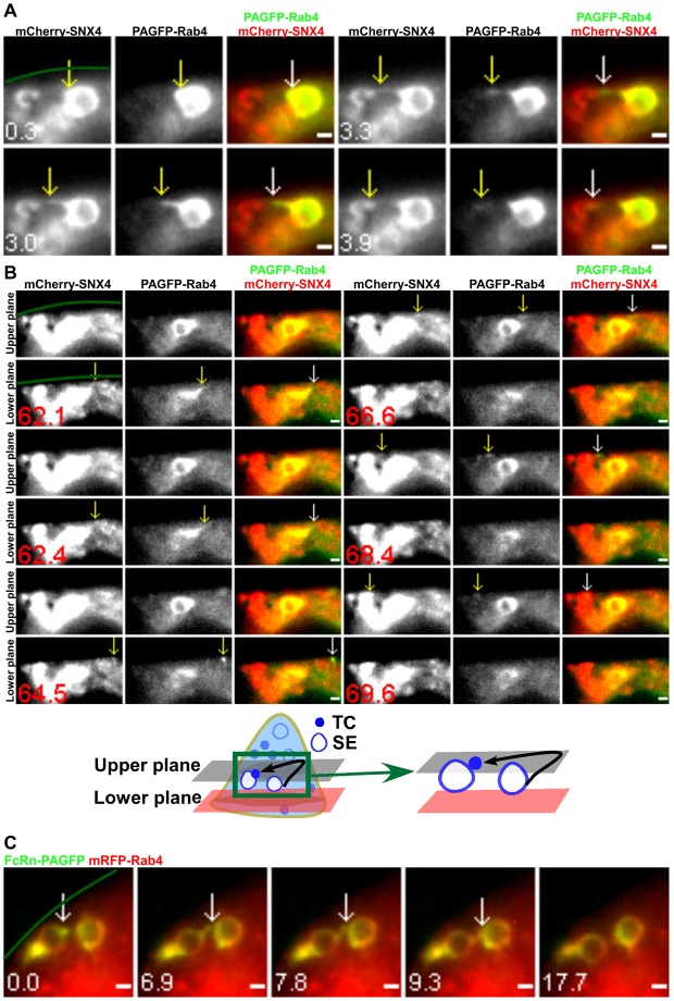
Interendosomal transfer TCs have associated SNX4/Rab4. HMEC-1 cells were cotransfected with mCherry–SNX4/PAGFP–Rab4 (A,B) or mRFP–Rab4/β2m/FcRn–PAGFP (C). PAGFP in individual sorting endosomes (SEs) was photoactivated. Scale bars: 1 µm. Green lines demarcate the plasma membrane. Images show selected frames from a dataset continuously acquired at a frame rate of 3 frames per second. (A) A SNX4+Rab4+ TC extends from a sorting endosome (0.3–3.3 s), segregates and subsequently merges with another sorting endosome (3.9 s). (B) Selected frames from supplementary material Movie 6. A second SNX4+Rab4+ TC leaves the same ‘donor’ sorting endosome as in A in the lower plane (62.1 s), moves rightwards at 64.5 s, changes direction and moves leftwards at 66.6 s. It merges with the ‘acceptor’ sorting endosome in the upper plane (69.6 s) (shown schematically below). (C) Two Rab4+ sorting endosomes were photoactivated sequentially. A FcRn+Rab4+ TC extends from one sorting endosome at 0 s, merges with the second sorting endosome (6.9–7.8 s) and subsequently segregates from the original endosome.

