Fig. 1.
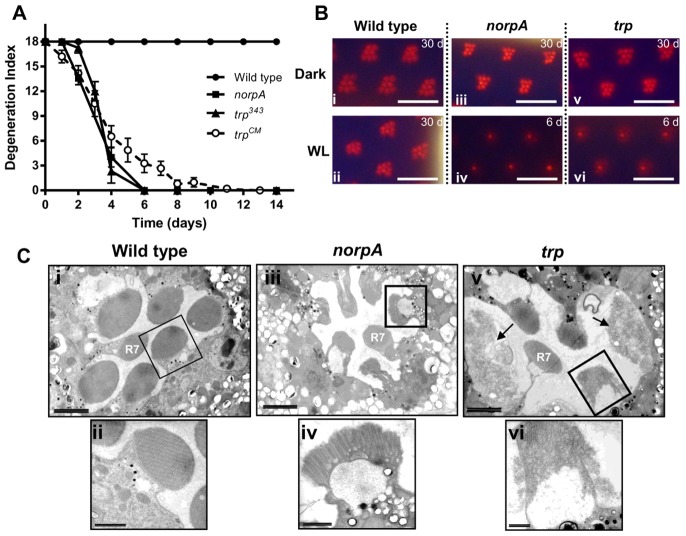
Retinal degeneration under white light. (A) Time course of retinal degeneration in wild type, norpAP24, and two independent trp mutants (trp343 and trpCM) under continuous white light (n = 5–7, means ± s.e.m.). (B) Representative optical neutralisation images of wild-type and mutant retinae after the indicated number of days (d) under continuous white light (WL) or darkness (Dark). (i,ii) Wild-type strain, Oregon R; (iii,iv) norpAP24; (v,vi) trp343 mutant. Scale bar: ∼16 µm. (C) Transmission electron microscopy (i,ii) Wild-type retina shows normal morphology after 5 days continuous white illumination. (iii,iv) norpAP24 retina exposed to white light for 5 days has short microvilli, but microvillar structure is still largely intact; (v,vi) trpCM retina exposed to white light for 5 days shows highly disintegrated microvilli giving a frothy appearance (black arrow). The rhabdomere of the UV-sensitive central photoreceptor (R7) in both mutants is spared from degeneration. Scale bars: ∼2 µm (Ci,iii,v); ∼500 nm (Cii,iv,vi).

