Fig. 5.
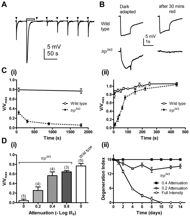
PtdIns(4,5)P2 depletion and retinal degeneration in trp mutants. (A) ERG recording from a trp mutant using a standard ERG set up. trp mutants show response inactivation to 200 ms dim test flashes (arrowheads) after prolonged illumination of 30 s (bar), reflecting PtdIns(4,5)P2 depletion (Hardie et al., 2001). The response gradually recovered, representing regeneration of PtdIns(4,5)P2. (B,C) PtdIns(4,5)P2 depletion in trp mutants under degenerative condition. (B) Representative traces from a wild type, and a trp mutant before (dark-adapted) and ∼5 s after exposure to the red LED in a light box for 30 minutes. (Ci) ERG amplitude response to a test flash as a function of duration of exposure to red LED, normalized to dark-adapted response prior to exposure (V/Vmax). 30 mins exposure caused near complete response inactivation (PtdIns(4,5)P2 depletion) in trp mutants but not in wild-type flies. (Cii) Response recovery after 30 mins of red light exposure (n = 5, means ± s.e.m.). (D) Intensity dependence of response inactivation (V/Vmax), reflecting PtdIns(4,5)P2 depletion (i), and retinal degeneration (ii) in trp mutants. Severity of PtdIns(4,5)P2 depletion (means ± s.e.m., n indicated in brackets) and retinal degeneration in trp (n≥7, means ± s.e.m.) had similar intensity dependence.

