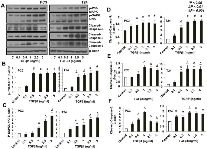
Figure 3. TGF β1 increased phosphorylation of p38 MAPK and SAPK/JNK and enhanced expression of cleaved caspases in PC3 and T24 cells.
(A) Western blots showing increased phosphorylation of p38-MAPK and SAPK/JNK, as well as increased expression of cleaved caspase-9, cleaved caspase-8, and cleaved caspase-3 after 24 h treatment with TGFβ1 (0.1, 0.5, 1, 2.5, and 5 ng/ml) compared to control (DMEM) (B–F) Bar graphs showing band-densitometry analysis of phoshorylations of p38 MAPK, SAPK and expression of cleaved caspase-9, cleaved caspase-8 and cleaved caspase-3, respectively, with TGFβ1 treatment as mentioned above, normalized to β-actin.

