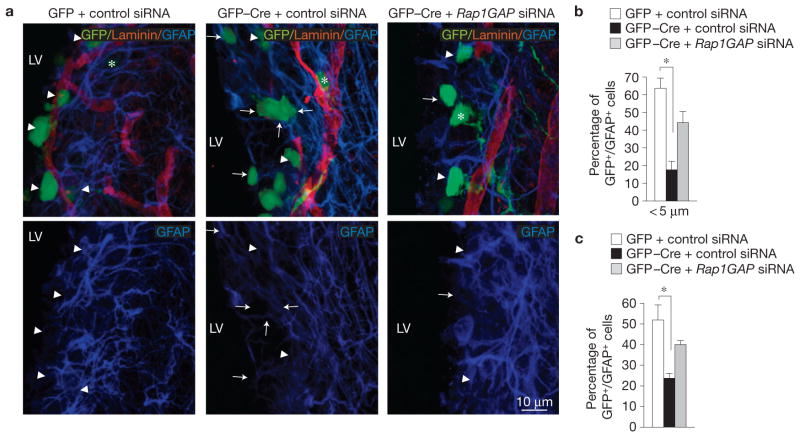
Figure 8.
Id proteins are required for adhesion of NSCs to the vasculature in the postnatal SVZ. (a) In vivo electroporation of MSCV–GFP or MSCV–GFP–Cre plasmid together with control or Rap1GAP siRNA into P5 mouse cortices, followed by immunostaining five days later. Cortical sections were stained for GFP to identify successfully transduced cells, GFAP (blue) to label NSCs and laminin (red) to define blood vessel surface. Arrowheads and arrows indicate GFP–GFAP-positive cells that lie <5 μm or >5 μm from laminin-positive structures, respectively. Asterisks indicate GFP-positive cells that are negative for GFAP. LV, lateral ventricle. (b), Quantification of GFP–GFAP-positive cells <5 μm from laminin+ structures. The asterisk indicates statistical significance. (c) Quantification of GFP–GFAP-positive cells. Results represent the means ± s.d.; n = 6 from 3 electroporated brains per group from two independent experiments. The asterisk indicates statistical significance. P = 0.0024 for b; P = 0.009 for c.

