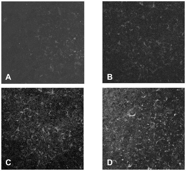
Figure 3.
Under normal, resting, conditions, the microvascular brain endothelial cells (MBEC) had minimal expression of the inflammatory markers VCAM-1 (A) or E-selectin (B). When the lower well (astrocyte compartment) was exposed to 100 Units/ml TNF-α for 24hrs the levels of both of these markers was increased on the apical surface of MBECs (C, VCAM-1; D, E-selectin). (Original magnification x20).

