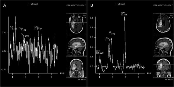
Fig. 3.
MR spectroscopic imaging of patient shown in Fig. 1 A with glioblastoma in temporal lobe treated with surgical resection. (A) Metabolite spectra corresponding to voxels selected inside the lesion. (B) Metabolite spectra corresponding to voxels selected inside the peri-lesional brain tissue. The lack of elevated choline (Cho) peak inside the lesion suggested treatment necrosis; however, histopathological verification proved it to be tumor recurrence.

