
Figure 3.
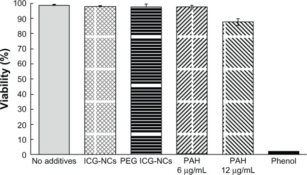
Viability of human dermal vascular endothelial cells after 24 hours’ incubation in media containing one of the following additives: uncoated indocyanine green-doped nanocapsules (ICG-NCs; 4.6 μg/mL), indocyanine green-doped polyethylene glycol-coated nanocapsules (PEG ICG-NCs, 4.5 μg/mL), and polyallylamine hydrochloride (PAH) at two different concentrations.
Notes: Cells incubated in media with no additive constituted the negative control, while cells incubated in media with phenol constituted the positive control. Each bar represents the average of three experiments. Error bars represent one standard deviation. Using statistics to analyze the results for ICG-NCs and PAH (6 μg/mL) yielded P values > 0.1 when compared with the negative control and P values < 10−4 when compared with the positive control. Therefore, these three tests indicated lack of adverse effects on cell viability. The same analysis for PAH at 12 μg/mL (2.6 times greater than the concentration administered in in vivo studies) yielded P values < 10−3 when compared with the negative control and P values < 10−4 when compared with the positive control.