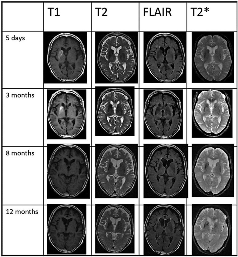
Fig. 1.
MRI. T1-weighted images revealed hyperintensity in the entire right striatum (putamen, caudate head and globus pallidus) at day 5 and at 3 months. The right striatum was slightly hypointense in T2-weighted and FLAIR images. T2*-weighted images demonstrated a low-intensity signal in the striatum predominantly on the right side at 3, 8 and 12 months. Atrophy of the right striatum was observed at 8 and 12 months. MRI was performed at the following facilities: day 5 (Suiseikai Kajikawa Hospital), 3 months (Hiroshima University School of Medicine), 8 and 12 months (Hiroshima General Rehabilitation Center).

