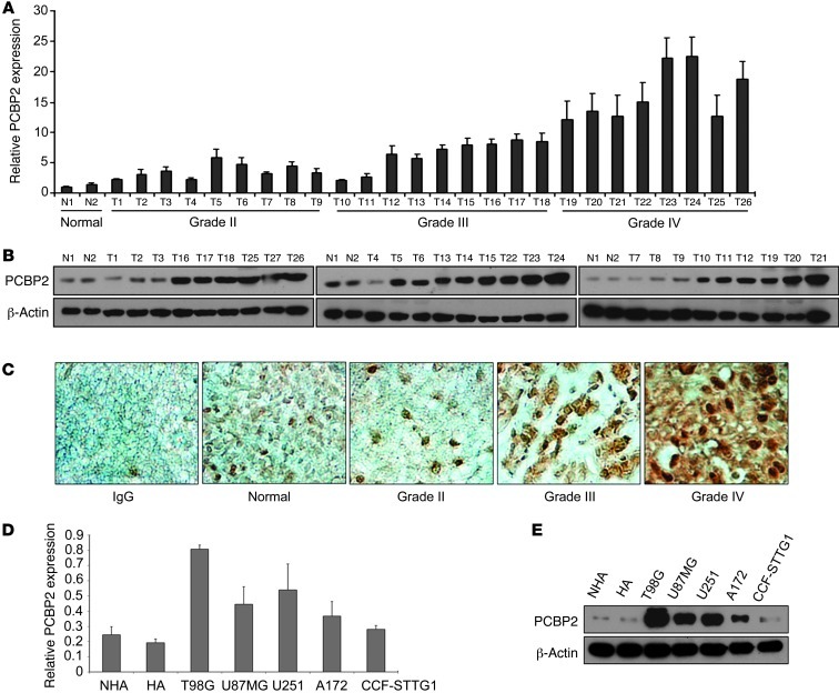
Figure 1. PCBP2 is upregulated in glioma tissues and cell lines compared with normal brain tissues and normal human astrocytes.
(A) Real-time PCR analysis of PCBP2 in 2 normal brain tissues (N1, N2) and 26 glioma tissues (T1–T9 grade II, T10–T18 grade III, and T19–T26 grade IV). Columns represent means and bars represent SD. (B) Representative Western blot showing PCBP2 protein levels in 2 normal brain tissues and 27 glioma tissues. β-Actin was used as a loading control. (C) Immunohistochemical staining of PCBP2 in glioma (Grade II, Grade III, Grade IV) and pericarcinous tissues (Normal) using anti-human PCBP2 antibodies. There was no staining with rabbit IgG. Original magnification, ×400. (D) Real-time PCR analysis of PCBP2 in 2 normal human astrocyte cell lines (NHA and HA) and 5 glioma cell lines (T98G, A172, U251, U87MG, and CCF-STTG1). (E) Representative Western blot showing PCBP2 protein levels in 2 NHA cell lines and the 5 indicated glioma cell lines.

