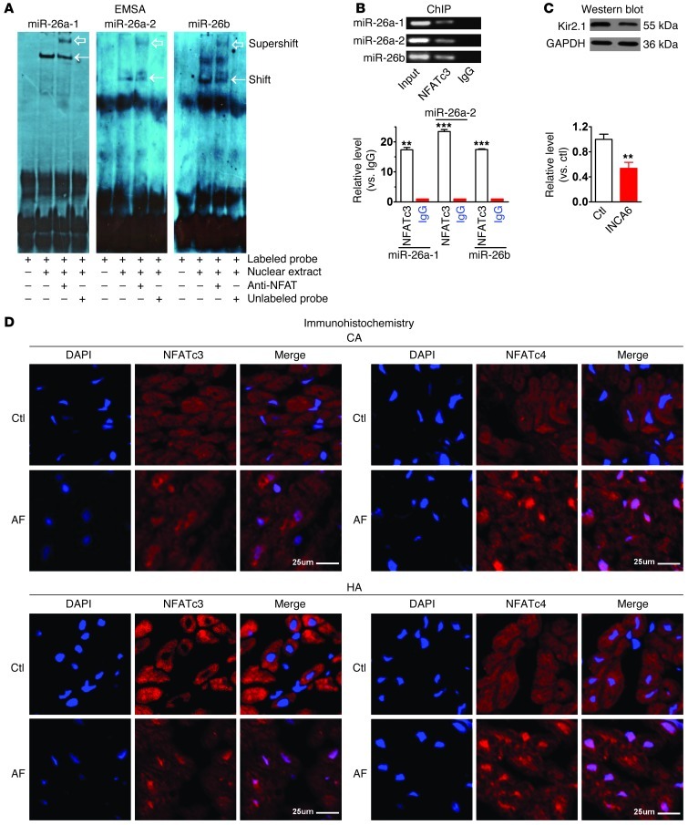
Figure 7. NFAT-dependent regulation of miR-26 in AF.
(A) EMSA to test in vitro binding of NFATc3 to its cis-acting element in the promoter region of human host genes for miR-26a-1, miR-26a-2, and miR-26b. Note shifted band representing protein-DNA binding (arrows), abolished with excess nonlabeled probe. Supershifted band (hollow arrows) represents NFATc3-cis-acting element binding in presence of anti-NFATc3 antibody. (B) ChIP testing in vivo binding of NFAT to human host genes of miR-26a-1, miR-26a-2, and miR-26b. Top: PCR products of 5′ flanking region encompassing NFAT-binding sites following immunoprecipitation with anti-NFATc3 antibody. Bottom: host gene binding measured by qPCR following ChIP, expressed as fold-changes over IgG-negative control. Input: genomic DNA without immunoprecipitation. **P < 0.01, ***P < 0.001 vs. IgG; n = 3/group. (C) Western blot showing Kir2.1 downregulation by INCA6 (100 nM). **P < 0.01 vs. control; n = 5/group. Values in B and C are mean ± SEM. (D) Immunohistochemical images showing nuclear translocation of NFATc3 and NFATc4 in atrial samples from AF dogs (CA) and patients (HA). Blue, nuclear staining (DAPI); red, NFAT staining; violet, NFAT nuclear localization in merged images. Findings similar to those in D were obtained in 3 subjects per group. Scale bars: 25 μm.

