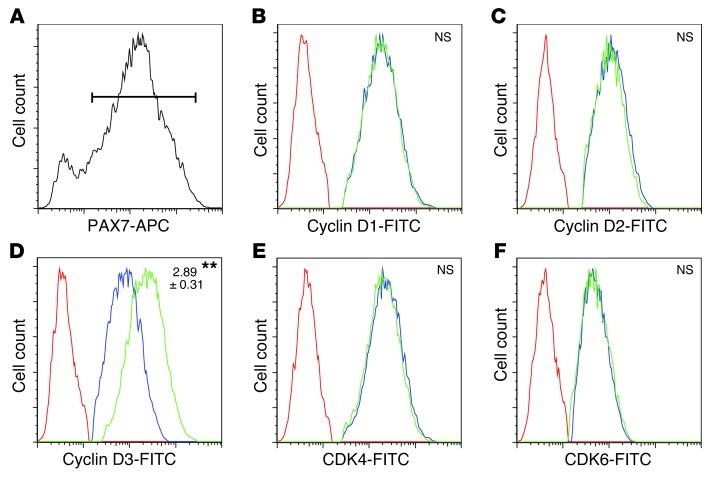
Figure 7. MKP-5 deficiency increases the expression of cyclin D3 in SCs.
Eight-week-old male mouse hind limb muscles were damaged by CTX, and 48 hours later muscles were dissected and digested to isolate cells for flow cytometry. Gated (PAX7+) cells (A) were analyzed for cyclin D1 (B), cyclin D2 (C), cyclin D3 (D), CDK4 (E), and CDK6 (F). No stain control (red); Mkp5+/+ (blue); Mkp5–/– (green). Data represent 3 independent experiments with 2 mice from each genotype in each independent experiment. Protein content was evaluated by measuring the median fluorescence intensity and expressed as fold change relative to Mkp5+/+. **P < 0.01 compared with Mkp5+/+ mice. NS, not statistically significant.

