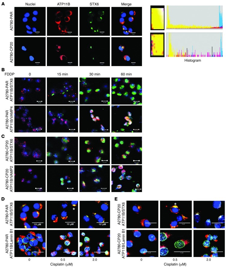
Figure 5. Subcellular localization of ATP11B in cisplatin-sensitive and cisplatin-resistant cells.
(A) Immunofluorescence staining using antibodies against ATP11B and specific cell markers showed the ATP11B signal mostly in the TGN colocalizing with STX6 (a TGN marker) in A2780-PAR and A2680-CP20 cells (green, STX6; red, ATP11B; blue, nuclei). Higher-magnification images (yellow boxes) from merged images show colocalization of ATP11B and STX6. Color histograms from these areas are included. (B) In both cell lines, FDDP, STX6, and ATP11B colocalized at the TGN at different times of FDDP exposure (15–60 minutes) (green, FDDP; red, ATP11B; blue, STX6). (C) Colocalization of FDDP, VAMP4 (vesicular transport marker), and ATP11B during FDDP exposure (green, FDDP; red, ATP11B; blue, VAMP4) in A2780-PAR and A2780-CP20 cells. Representative confocal images after 0, 15, 30, and 60 minutes of FDDP exposure. (D) Confocal images from A2780-PAR and (E) A2780-CP20 cells in the absence and presence of nonfluorescent cisplatin (0–2 μM), immunostained for the detection of ATP11B/ STX6 and ATP11B/lamin B1 (nuclear marker). In both cell lines colocalization between ATP11B and STX6 was stronger in the presence of cisplatin (asterisks). ATP11B was frequently observed in nuclei from A2780-PAR cells colocalizing with lamin B1 (green, STX6, lamin B1; red, ATP11B; blue, nuclei). Original magnification, ×200; scale bars: 5 μM (A); 10 μM (B–E).

