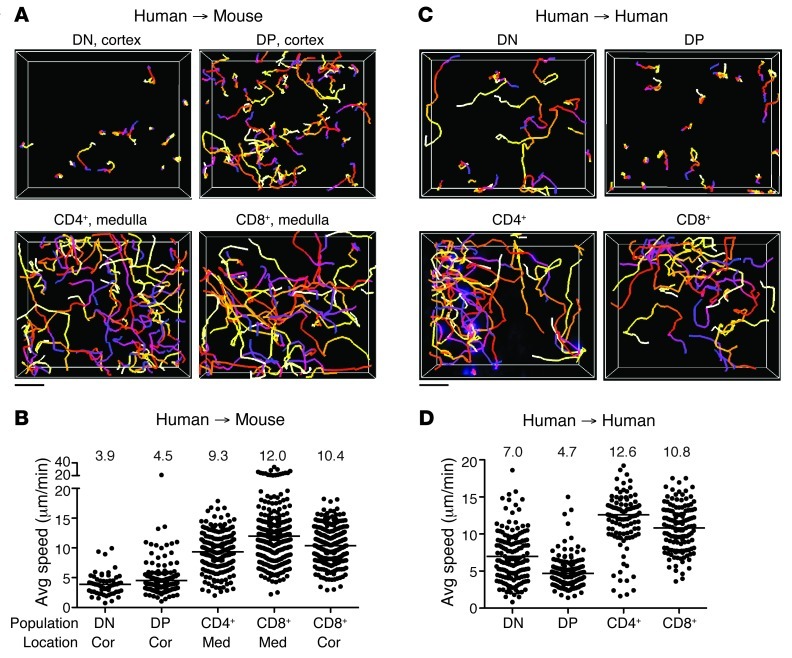
Figure 2. Human and mouse thymic slices support human thymocyte migration.
(A) Cell tracks from representative 2-photon time-lapse datasets of human thymocyte subsets on CD11c-YFP thymic slices (20-minute movies). (B) Average speed of human thymocytes on CD11c-YFP thymic slices. CD4+ and CD8+ SP movies were acquired in the medulla (med) or cortex (cor); DN and DP movies were acquired in the cortex. n = 57 tracks (DN); 178 tracks (DP); 207 tracks (CD4+); 274 tracks (CD8+ in medulla); 279 tracks (CD8+ in cortex). (C) Cell tracks from representative 2-photon time-lapse movies of human thymocyte subsets on human thymic slices (30-minute movies). (D) Average speed of human thymocytes on human thymic slices. n = 151 tracks (DN); 187 tracks (DP); 107 tracks (CD4+); 135 tracks (CD8+). (A and C) Tracks are color-coded to indicate passage of time (blue→red→yellow→white). (B and D) Symbols represent average speed of individual tracked cells; lines represent average value of compiled track averages for each population. Scale bars: 30 μm.

