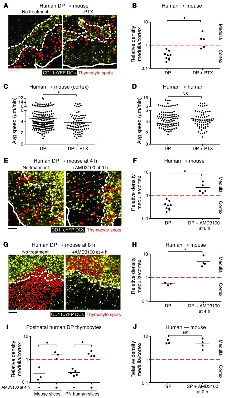
Figure 6. CXCR4 directs the appropriate localization of human DP thymocytes.
(A and B) Representative cryosections (A) and relative density (B) of untreated (from Figure 1D) and PTX-treated (n = 358) fetal DP thymocytes on CD11c-YFP slices. (C and D) Average speed of (C) untreated (from Figure 2B) or PTX-treated (n = 80) fetal DP cells in the cortex of CD11c-YFP slices and on (D) human fetal thymic slices. n = 97 (DP); 73 (DP+PTX). (E and F) Fetal DP thymocytes were incubated with AMD3100 at the start of culture, and their location was determined after 4 hours. Representative cryosections (E) and relative density (F) of untreated (from Figure 1D) or AMD3100-treated (n = 2,358) fetal DP thymocytes on CD11c-YFP slices. (G and H) Fetal DP thymocytes were overlaid on CD11c-YFP slices for 4 hours, washed, then treated with AMD3100 for another 4 hours. Representative cryosections (G) and relative density (H) of untreated (n = 5,282) or AMD3100-treated (n = 1,530) fetal DP thymocytes on CD11c-YFP slices. (I) Postnatal (PN) DP thymocytes were overlaid onto CD11c-YFP or human postnatal slices for 4 hours, washed, then treated or not with AMD3100 for another 4 hours. n = 1,006 (mouse DP); 2,221 (mouse DP+AMD3100); 2,752 (postnatal human DP); 1,308 (postnatal human DP+AMD3100). (J) Relative density of untreated (n = 1,392) or AMD3100-treated (n = 1,605) fetal CD4+ and CD8+ SP thymocytes on CD11c-YFP slices. Data were compiled from at least 3 tissue sections from at least 2 independent experiments. (A, E, and G) Solid and dashed outlines as in Figure 1. (C and D) Symbols and lines as in Figure 2. (B, F, and H–J) Each dot represents quantification of 1 tissue section. Scale bars: 100 μm. *P < 0.05.

