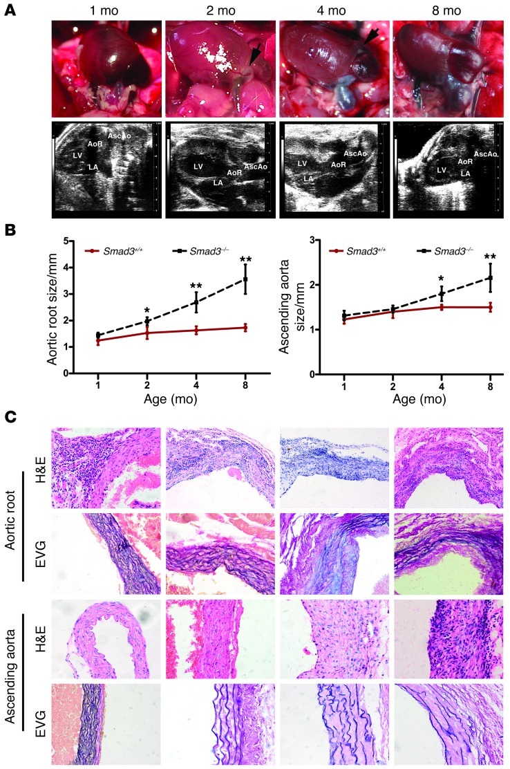
Figure 2. Smad3–/– mice undergo progressive aortic root and ascending aorta dilation.
(A) Representative photographs and ultrasound imaging of the aortic root and ascending aorta in Smad3–/– mice at different ages. Arrows in the photographs of 2-month-old mice identify areas of neovascularization. (B) Aortic root and ascending aortic diameter, measured by echocardiography, at different ages in Smad3+/+ (n = 9/time points) and Smad3–/– (n = 7/time points) mice. *P < 0.01; **P < 0.001, Smad3–/– versus Smad3+/+ at the same age. (C) H&E staining showed inflammatory cell infiltration in the aortic roots and ascending aortas of Smad3–/– mice (n = 12/time points) at different ages. EVG staining showed medial elastin degradation in the aortic roots and ascending aortas of Smad3–/– mice (n = 11/time points) at different ages. Original magnification, ×200 (C).

