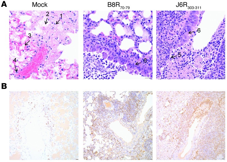
Figure 7. Epitope-specific TCD8 infiltrated infected lungs and protected them from severe damage.
(A) Hematoxylin and eosin stain of infected lungs from nonvaccinated (left) and vaccinated (middle, right) mice. Severe pathology shows wide spread necrosis (no. 1), extensive fibrin deposition (no. 2), edema (no. 3), and vasculitis (no. 4). Moderate damage shows necrosis limited to airway epithelium (no. 5) and signs of regeneration (epithelial hyperplasia; no. 6). (B) Anti-CD3 staining of the infected lungs. Mice vaccinated with B8R70–79 and J6R303–311 (see Figure 5) show prominent T cell infiltration around airways, which is present in low numbers in the lungs of mock-vaccinated mice. Scale bar: 25 μM.

