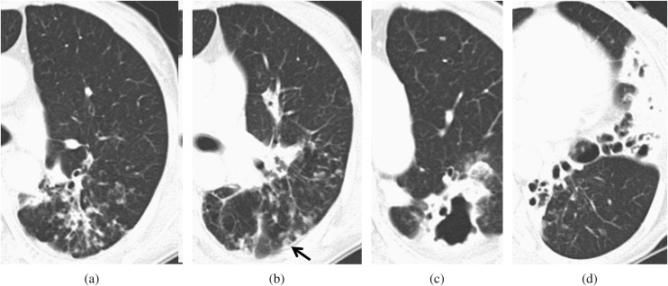
Figure 2.
A 56-year-old female on steroid therapy for 1 year. The total disease extent was 10. (a, b) CT scan shows bronchiectasis with ill-defined nodules in the superior division of the left upper lobe. A cavity (black arrow) in an ill-defined nodule was also seen. The bronchiectasis score was 4. (c, d) CT scan shows a large opacity >2 cm, with a cavity and bronchiectasis in the left lower lobe. The bronchiectasis severity was 3. The bronchiectasis score was 9.

