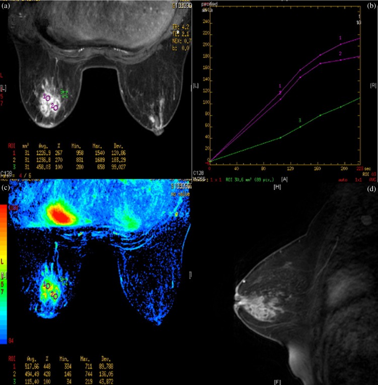
Figure 6.
Female, 47 years old, acute/chronic mastitis. Enhanced MR image showed multiple, contiguous, clustered, rim-like enhancements in the left breast (a); Type 1 time–signal intensity curves were obtained (b, c) and the sagittal MR image showed a non-mass-like enhanced lesion directed to the nipple along the breast ducts (d).

