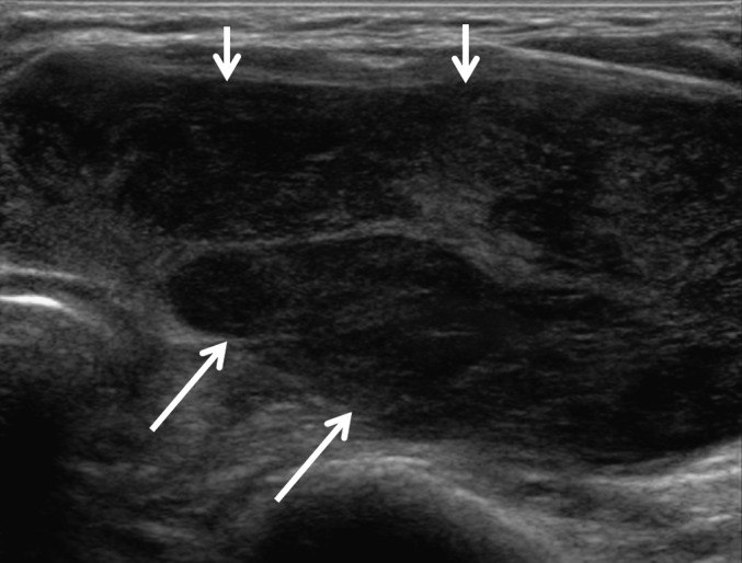
Figure 3.

Group C. Suspicious for primary lymphoma seen on ultrasonography. An 82-year-old female had diffuse heterogeneous hypoechoic parenchyma (arrows) with intervening echogenic septa-like structures in the left thyroid gland. She had a history of a rapidly growing mass. The cytological result showed the differential diagnosis of florid lymphoid hyperplasia and non-Hodgkin’s lymphoma and was inconclusive. Ultrasonography-guided core needle biopsy diagnosed diffuse large B-cell lymphoma.
