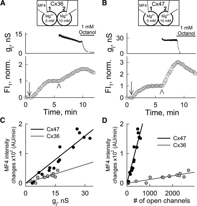
Figure 5.
Permeation of Cx36 or Cx47 GJ channels by Mg2+ ions. A, B, Mag-fluo-4 (MF4) fluorescence intensity measured in cell-1 (FI1) of HeLa Cx36-EGFP (A) and HeLa Cx47-EGFP (B) cell pairs increased after opening the patch in cell-1 (arrow). After FI1 reached a plateau (normalized to this value), pipette-2 was opened (arrowhead) and FI1 again increased. Pipette-1 contained 50 μm MF4 and zero MgCl2, and pipette-2 contained 10 mm MgCl2 (top diagram). The gj measurements were started after patch opening in cell-2 (top). FI1 and gj decreased after bath application of the GJ blocker octanol (1 mm); decrease in FI1 is ascribable to loss of Mg2+ into pipette-1. C, D, Rates of FI1 changes in HeLa Cx36-EGFP (gray; n = 12) and HeLa Cx47-EGFP (black; n = 15) cell pairs measured after patch opening in cell-2 and plotted over gj (C) or the calculated number of open channels (D). Gray and black lines are linear regressions for Cx36-EGFP (R2 = 0.71) and Cx47-EGFP (R2 = 0.9) data, respectively.

