
Fig. 2.
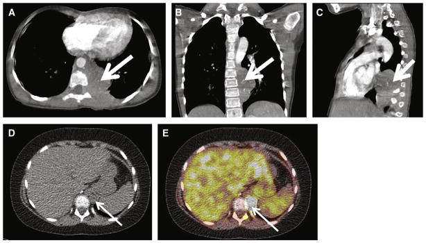
Supradiaphragmatic PGL in a 9-year-old SDHB+ girl with a 4-year history of heat intolerance and malaise. A–C, Axial, coronal, and sagittal contrast-enhanced CT images, respectively, demonstrate a 5.9 × 3.5 × 6.1-cm left posterior mediastinal mass (white arrow). Invasion of the left T8 neural foramen is demonstrated on the axial image (A, white arrowhead). Arterial phase CT attenuation of this mass was 52 HU; portal venous phase CT attenuation of this mass was 88 HU. The patient underwent thoracotomy and resection of the posterior mediastinal mass, confirming PGL. D–E, at 15 months after resection, FDG PET-CT images demonstrate a new 1.5-cm retrocrural metastasis, with maximal SUV of 34 (white arrow) as well as pleural based masses (not shown), consistent with metastases.