Fig. 3.
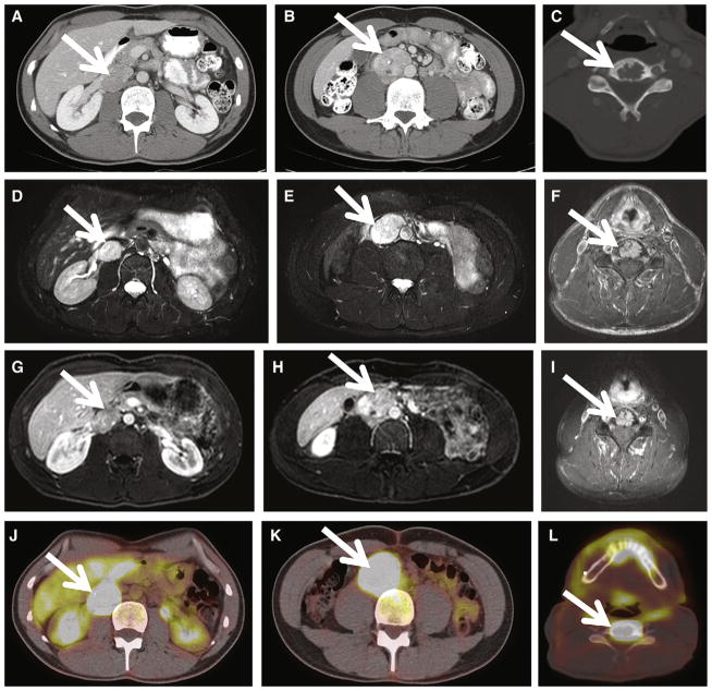
Shown is a 34-year old SDHB+ man with history of hypertension, tachycardia, and heat intolerance with multifocal and metastatic PGLs. A, Axial contrast CT at the level of the renal veins demonstrates a 3-cm enhancing right renal hilar mass, consistent with extra-adrenal PGL(white arrow). Portal venous phase CT attenuation of this mass was 139 HU. B, Axial contrast CT inferior to the renal vein confluence demonstrates a 4-cm right para-aortic retroperitoneal mass (white arrow), consistent with PGL. Portal venous phase CT attenuation of this mass was 139 HU. C, Osteolytic metastasis to the C5 vertebral body (white arrow). D–E, Axial T2-weighted images demonstrate homogenous T2 hyperintensity of patient’s right retroperitoneal PGLs. T2 signal is hypointense to CSF but hyperintense to liver and spleen (group 2 classification according to criteria proposed by Jacques et al13). F, FSE T2-weighted MRI demonstrates T2 hyperintense C5 metastasis (white arrow). G–I, Axial postcontrast MRIs demonstrate enhancing right retroperitoneal PGLs and C5 metastasis, respectively (white arrow). J–L, 18F-FDG-PET CT images demonstrate hypermetabolic foci corresponding to patient’s right retroperitoneal PGLs and C5 osseous metastasis with maximal SUVs of 22, 26, and 42, respectively. The patient underwent exploratory laparotomy, resection of a right renal hilar extra-adrenal PGL, and interaortocaval extra-adrenal PGL proximal to the aortic bifurcation.

