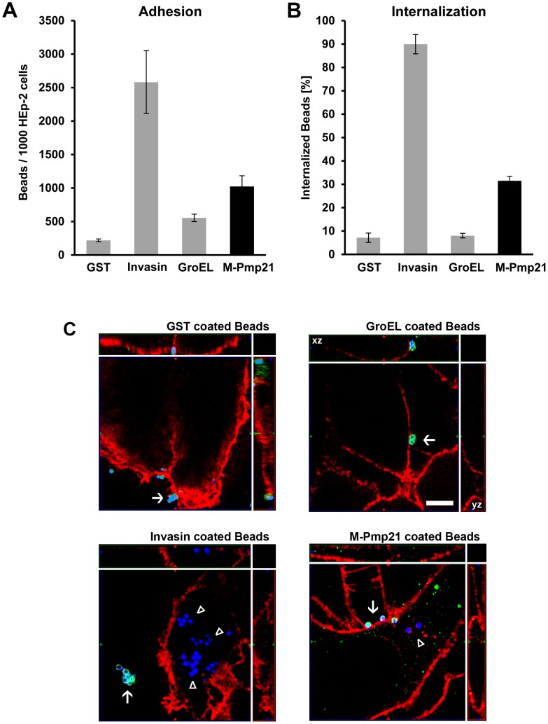
Figure 1. Pmp21-coated beads are taken up by mammalian cells.
(A) Green fluorescent beads coated with recombinant GST, invasin, GroEL1 or M-Pmp21 were incubated in 5-fold excess with HEp-2 cells for 1 h at 4°C, and the numbers of beads found on 1000 HEp-2 cells were counted (n = 3). (B) Internalization of M-Pmp21-coated beads. HEp-2 cells were incubated with beads as above at 37°C for 4 h, and washed with PBS to remove unattached beads. Attached beads were stained with specific antibodies without cell permeabilization (see Figure S1), and the numbers of attached (red) and internalized (green) beads on/in samples of 1000 cells were counted (n = 3). (C) Confocal spinning-disk images of internalized beads (for the GroEL image the xz and yz plain projections of the whole field are marked). HEp-2 cells were incubated for 4 h at 37°C with blue fluorescent beads coated with GST, invasin, GroEL1 or M-Pmp21. External beads were stained with protein specific antibodies (green). Internalized beads are not accessible to the antibody and appear blue. Cell boundaries were stained with Wheat Germ Agglutinin-Alexa594 (red). External beads are marked by white arrows, internalized beads by white triangles. Bar 5 µm.

