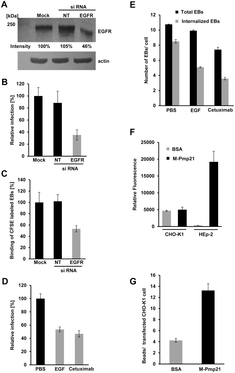
Figure 4. EGFR is required for internalization of C. pneumoniae.
(A–C) HeLa229 cells were mock-transfected (Mock), or transfected with a non-targeting siRNA (NT) or an EGFR-targeting siRNA for 24 h. (A) Relative EGFR levels were determined using Scion Image software after immunoblot analysis with anti-EGFR. Actin served as loading control. (B) HeLa229 cells transfected for 24 h were infected with C. pneumoniae GiD (MOI 1) for 48 h. Inclusion formation was evaluated by indirect immunofluorescence with an FITC-conjugated antibody against chlamydial LPS. The number of inclusions in mock-transfected cells was set to 100% (n = 4). (C) C. pneumoniae EBs labeled with CFSE were added (MOI 10) to transfected HeLa229 cells for 1 h at 37°C. Cells were then detached from the substrate and fixed with formaldehyde, and adhesion was measured by flow cytometry. The mean fluorescence of EBs bound to mock-transfected cells was set to 100% (n = 3). (D) Pretreatment of confluent HEp-2 monolayers with recombinant EGF or Cetuximab for 2 h inhibits subsequent infection (n = 4) (MOI 1). (E) Pretreatment of HEp-2 cells with rEGF or Cetuximab for 2 h inhibits subsequent internalization of C. pneumoniae EBs (MOI 1). Cells were exposed to EBs for 2 h, then fixed and stained with anti-Pmp21 and DAPI without permeabilization. Numbers of internalized EBs (inaccessible to anti-Pmp21 antibody) were determined by subtracting the number of external EBs (visualized with anti-Pmp21) from the total number of EBs (DAPI stain) in each cell (n = 5). (F) rM-Pmp21-coated beads adhere to HEp-2 but not CHO-K1 cells. A five-fold excess of green fluorescent beads coupled to BSA or rM-Pmp21 were incubated with CHO-K1 and HEp-2 cells for 1 h at 37°C. Unbound beads were removed by washing with PBS, and cells bearing attached beads were analyzed by flow cytometry. The mean fluorescence values for the samples analyzed (n = 4) are shown. (G) rM-Pmp21-coated beads attach to EGFR-expressing CHO-K1 cells. CHO-K1 cells were transfected with EGFR-mCherry for 24 h, incubated with a five-fold excess of green fluorescent beads coated with BSA or rM-Pmp21, and the numbers of beads attached to transfected cells were determined (n = 3).

