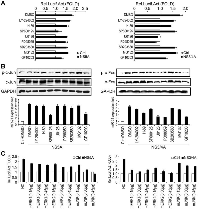
Figure 3. Investigation of the roles of ERK, JNK, and PKC in the regulation of miR-21 expression upon HCV infection.
(A) Huh7 cells were co-transfected with miPPR21 and pCMV-NS5A (left panel) or pCMV-NS3/4A (right panel) for 24 h, and then signal pathway specific inhibitors (20 µM each) were then added, as indicated. The cells were lysed and luciferase activity was measured. (B) Cells were transfected with pCMV-NS5A (left panel) or pCMV-NS3/4A (right panel) for 24 h, and then treated with the signal pathway inhibitors (20 µM each) as indicated. The phosphorylation and total protein levels of c-Jun (left panel) and c-Fos (right panel) were determined by Western blot (upper panel), and miR-21 expression was measured by qPCR (lower panel). (C) Huh7 cells were co-transfected with miPPR21 and dominant-negative mutants of ERK1 (mERK1), ERK2 (mERK2), JNK (mJNK) or control vectors at different concentrations, as indicated and the resultant luciferase activities were measured. All experiments were repeated at least three times with similar results. Bar graphs represent the means ± SD, n = 3.

