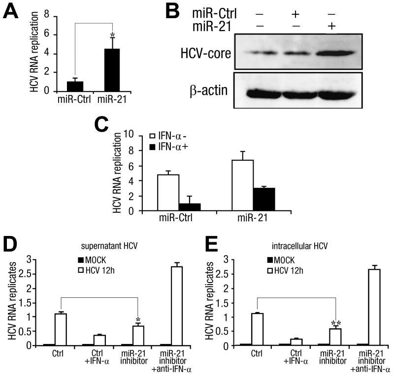
Figure 6. miR-21 stimulates HCV replication and attenuates the HCV response to IFN-α treatment.
(A) Huh7 hepatocytes were transfected with control RNA (miR-ctrl) or miR-21 mimics (final concentration, 50 nM). After 48 h, cells were infected with HCV (MOI = 1) for 2 h and washed before fresh medium was added. After 72 h, intracellular HCV RNA replicates were quantified by qPCR and normalized to the GAPDH internal control. (B) Huh7 hepatocytes were transfected as described in (A) and infected with HCV (MOI = 1) for 2 h. After 48 h, HCV core expression was analyzed by Western blot (top panel) using β-actin as a loading control (bottom panel). (C) Huh7 hepatocytes were transfected with miR-21 mimics or control RNA (miR-ctrl) (final concentration, 50 nM). After 48 h, cells were infected with HCV (MOI = 1) for 2 h and washed before adding fresh medium with or without recombinant human IFN-α (100 U/ml). After 72 h, intracellular HCV RNA replicates were quantified by qPCR. (D and E) Huh7 hepatocytes were transfected with miR-21 inhibitors or control inhibitor (ctrl) (final concentration, 50 nM). After 48 h, cells were infected with HCV (MOI = 1) for 2 h and washed before adding fresh medium with or without recombinant human IFN-α (100 U/ml) or anti-IFN-α-neutralizing antibody (100 neutralizing units/ml) as indicated. After 72 h, RNA was isolated from the cell culture medium, and supernatant HCV replicates (D) were measured by qPCR. Intracellular HCV RNA replicates (E) were quantified by qPCR using GAPDH as internal control. Data are presented as the means SD (n = 3) from one representative experiment. Similar results were obtained in three independent experiments. **, p<0.01; *, p<0.05.

