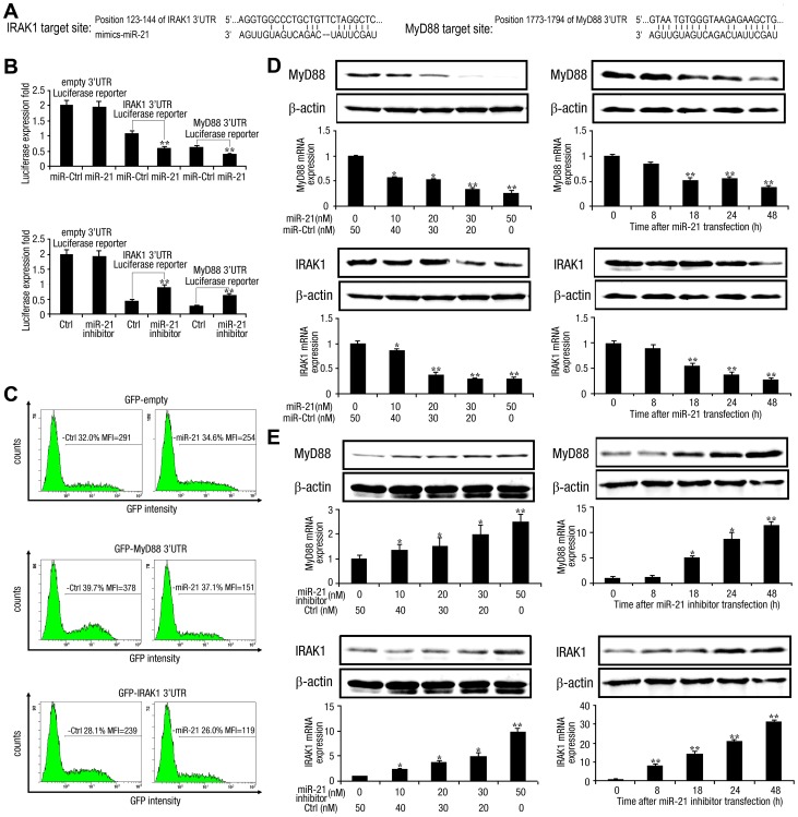
Figure 9. miR-21 targets human MyD88 and IRAK1.
(A) Sequence alignment of miR-21 and its binding sites in the 3′ UTRs of MyD88 and IRAK1, as predicted by RNA22 software. (B) Huh7 hepatocytes (1×10⁴) were co-transfected with pGL3-Basic, pGL3-MyD88 3′ UTR, or pGL3-IRAK1 3′ UTR firefly luciferase reporter plasmids (80 ng) and pRL-TK Renilla luciferase plasmid (40 ng), together with miR-21 mimics or control RNA, miR-21 inhibitor or control inhibitor (final concentration, 50 nM), as indicated. After 48 h, firefly luciferase activity was determined and normalized to Renilla luciferase activity. (C) HEK293 cells (1×10⁴) were co-transfected with GFP control, GFP-MyD88 3′ UTR, or GFP-IRAK1 3′ UTR plasmid (400 ng), together with miR-21 mimics or control RNA (final concentration, 50 nM), as indicated. After 48 h, GFP expression was analyzed by FACS, and the mean fluorescence intensity (MFI) of GFP was determined. (D and E) Huh7 hepatocytes (1×10⁶) were transfected with miR-21 mimics (D) or miR-21 inhibitor (E) at various concentrations for 48 h (left), or at 50 nM (final concentration) for the indicated time (right). MyD88 and IRAK1 protein levels were determined by Western blot and normalized to β-actin (top panel); MyD88 and IRAK1 mRNA levels were determined by qPCR and normalized to GAPDH (bottom panel). Data are presented as the means SD (n = 3) from one representative experiment. Similar results were obtained in three independent experiments. **, p<0.01; *, p<0.05.

