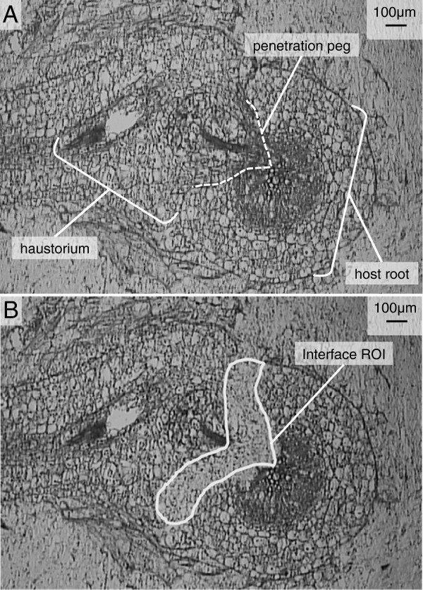
Figure 1.
Laser Microdissected Haustorium. LPCM allows highly tissue- and cell-specific harvest after histological identification of tissues or cells of interest. A) Representative 25 μm cross-section of T. versicolor haustorium on the host M. truncatula approximately 9 days post infestation, and prior to LPCM. The mature haustorium contains the xylem bridge that connects the parasite and host vasculature and is visible in the penetration peg. B) The same section after LCPM shows the cleared interface tissue from the user-defined region of interest (ROI). The flakes of tissue are catapulted by a photonic cloud resulting from pulses of laser light focused between the tissue and glass slide. Multiple pulses of laser light raster across the ROI causing tissue in the selected region to be catapulted and then captured in the adhesive coated cap of a 0.5 mL tube held by a robotic arm in very close proximity (< 0.5 mm) to the upper surface of the section affixed to the slide.

