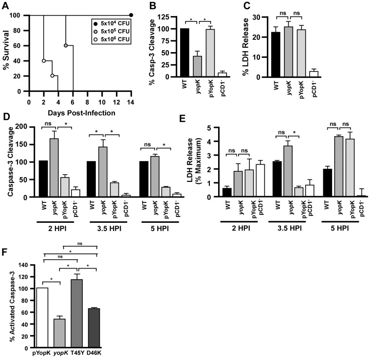
Figure 1. YopK modulates caspase-3 activation in vitro in Yersinia pestis infected macrophages.
A) Intravenous infection of C57BL/6 mice with the indicated doses of KIM D27 yopK for determination of 50% lethal dose (data shown are representative of two trials; n = 8–10 mice per group total). RAW 264.7 cells were infected with Y. pestis KIM D27, yopK, pYopK and CO92 pCD1−, (B) harvested after 3.5 hours and assayed for caspase-3 activation (shown are the mean values from four trials, each sample run in duplicate) or (C) harvested after 4 hours for release of lactate dehydrogenase (LDH) (shown are the mean values from nine trials, each sample run in duplicate). (D–E) MH-S cells, an alveolar macrophage cell line, were infected with KIM D27, yopK, pYopK, and KIM6- (pCD1−) and assayed for caspase-3 activation (D) and LDH release (E) at 2, 3.5, and 5 HPI, shown are the mean values from two (5 HPI), four (2 HPI) or six (3.5 HPI) trials, each sample run in duplicate. (F) RAW 264.7 macrophages were infected at an MOI of 20 for 3.5 hours with Y. pestis KIM D27yopK complemented with wild type yopK or the T45Y or D46K mutants; data shown are the mean values collected in 2–5 independent trials, with each sample run in duplicate. Caspase data are reported as a percentage of caspase activation by WT Y. pestis KIM D27; LDH data are reported as a percent of total LDH released following lysis of macrophages that were not infected. Data from each trial were evaluated for statistical significance using one-way ANOVA followed by the Tukey post-hoc test, *p<0.05, ns not significant.

