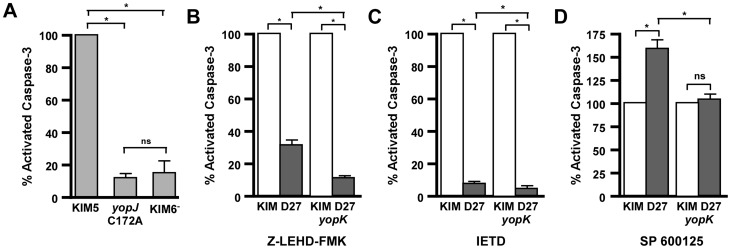
Figure 2. YopK contributes to caspase-3 cleavage through the extrinsic pathway.
A) RAW 264.7 macrophages were infected with KIM5, KIM5 yopJC172A, or KIM6− at an MOI of 20 and assayed for caspase-3 activity after 3.5 hours of infection. Data shown are the mean of four biological replicates, collected in two independent trials, and are shown as a percentage of caspase-3 activity from WT-infected cells. B–D) RAW 264.7 macrophages were left untreated (white bars) or treated with inhibitors (grey bars) for (B) caspase-9 (Z-LEHD-FMK), (C) caspase-8 (IETD), or (D) JNK (SP600125) and infected with either Y. pestis KIM D27 or KIM D27 yopK at an MOI of 20. After 3.5 hours of infection, cell pellets were analyzed for cleaved caspase-3. Data shown are the mean of four replicates, collected in two independent trials and are shown as percent of untreated for each strain. Statistical significance by one-way ANOVA followed by Tukey post-hoc test, *p<0.05, ns not significant.

